Home
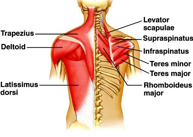
/ Back Muscles Anatomy Labeled - Labeled Human Anatomy Diagram Of Man S Arm Shoulder And Upper Back Stock Images Page Everypixel / The back anatomy includes the latissimus dorsi, trapezius, erector spinae, rhomboid, & teres major.
Back Muscles Anatomy Labeled - Labeled Human Anatomy Diagram Of Man S Arm Shoulder And Upper Back Stock Images Page Everypixel / The back anatomy includes the latissimus dorsi, trapezius, erector spinae, rhomboid, & teres major.
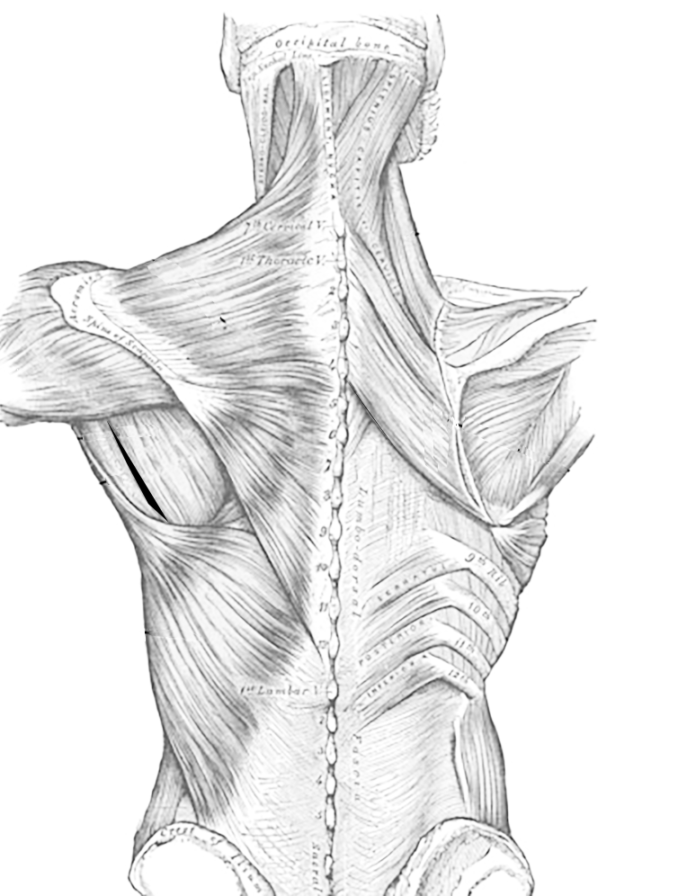
Back Muscles Anatomy Labeled - Labeled Human Anatomy Diagram Of Man S Arm Shoulder And Upper Back Stock Images Page Everypixel / The back anatomy includes the latissimus dorsi, trapezius, erector spinae, rhomboid, & teres major.. The muscles of the back are separated into extrinsic and intrinsic components, which are based on their function in movement and embryological origin. Learn about anatomy muscle labeling with free interactive flashcards. The general action of the back muscles allows movement in the head, shoulders, arms, and the spine they are also involved in movement of the ribs which allows for respiratory function. The muscles of the back are a group of strong, paired muscles that lie on the posterior aspect of the trunk. Muscles, connected to bones or internal organs and blood vessels, are in charge for movement.
Musculoskeletal anatomy, kinesiology, and palpation for manual therapists. The extrinsic back muscles are also referred to as secondary back muscles. They are divided into three groups, as shown below. Exercise of this organ system is critical to prevent. The muscular system is responsible for the movement of the human body.
Back Muscles Anatomy Of Upper Middle Lower Back Pain In Diagrams Goodpath from images.ctfassets.net This is my video about the muscles of the back. The extrinsic muscles that are associated with upper extremity and shoulder movement, and the intrinsic injuries of the intrinsic back muscles often occur while using improper lifting technique. Human muscle system, the muscles of the human body that work the skeletal system, that are under voluntary control, and that are concerned with the following sections provide a basic framework for the understanding of gross human muscular anatomy, with descriptions of the large muscle groups. Let's remember the back muscles. Choose from 500 different sets of flashcards about anatomy muscle labeling on quizlet. Neck muscle anatomy mri 12 photos of the neck muscle anatomy mri neck muscle anatomy images, neck muscle anatomy pictures, neck muscle anatomy posterior, neck muscle anatomy ultrasound, neck muscles anatomy radiology. The muscular system is responsible for the movement of the human body. The muscular system is responsible for movement in collaboration with the nervous system to form impulses for motion.
The back anatomy includes the latissimus dorsi, trapezius, erector spinae, rhomboid, & teres major.
Labels are a means of identifying a product or container through a piece of fabric, paper, metal or plastic film onto which information about them is printed. It inserts on the proximal end of the humerus and its primary. This is a table of skeletal muscles of the human anatomy. The muscular system is responsible for movement in collaboration with the nervous system to form impulses for motion. This article covers the anatomy of the deep muscles of the back, including their function, blood supply, innervation, origin and insertion. Muscle basics and cellular components, naming of the muscles, and cat. The muscles of the back are a group of strong, paired muscles that lie on the posterior aspect of the trunk. The extrinsic muscles that are associated with upper extremity and shoulder movement, and the intrinsic injuries of the intrinsic back muscles often occur while using improper lifting technique. Included are several layered views of the back muscles, the doral muscles, subclavius muscles, rhomboideus major and minor muscles, deltoid muscles and many more. The muscles of the back are separated into extrinsic and intrinsic components, which are based on their function in movement and embryological origin. These muscles give height and breadth to back. This is my video about the muscles of the back. Back muscles are divided into two specific groups:
The extrinsic muscles include the trapezius, latissimus dorsi, rhomboid major and minor, levator scapulae and the serratus posterior superior and. Labels are a means of identifying a product or container through a piece of fabric, paper, metal or plastic film onto which information about them is printed. Tutorials on the anatomy and actions of the back muscles, using interactive animations, diagrams, and illustrations. Topographically, the muscles in this group are classed along with the lateral torso wall and upper extremity, which is due to their location as well as their genetic development based on their embryological origin. The superficial back muscles are the muscles found just under the skin.
Muscles Of The Thoracic Region Dorsal Side from www.biologycorner.com There are around 650 skeletal muscles within the typical human body. This site was designed for students of anatomy and physiology. The muscular system is responsible for movement in collaboration with the nervous system to form impulses for motion. These muscles give height and breadth to back. Intermediate back muscles and c. The back muscles can be three types. They are divided into three groups, as shown below. Learn about these muscles, their locations there are several individual muscles within the back anatomy, and it's important to take a quick look at all of them to see how you can target them.
It inserts on the proximal end of the humerus and its primary.
Labels are a means of identifying a product or container through a piece of fabric, paper, metal or plastic film onto which information about them is printed. Neck muscle anatomy mri 12 photos of the neck muscle anatomy mri neck muscle anatomy images, neck muscle anatomy pictures, neck muscle anatomy posterior, neck muscle anatomy ultrasound, neck muscles anatomy radiology. Learn all about the muscles of the back in this 3d video anatomy tutorial. Anatomical diagram showing a back view of muscles in the human body. Learn anatomy faster and remember everything you learn. Microscopic anatomy of skeletal muscle. This quiz requires labeling, so it will test your knowledge on how to identify these muscles (latissimus dorsi, trapezius, deltoid, biceps brachii. Topographically, the muscles in this group are classed along with the lateral torso wall and upper extremity, which is due to their location as well as their genetic development based on their embryological origin. The extrinsic back muscles are also referred to as secondary back muscles. There are around 650 skeletal muscles within the typical human body. Their main function is contractibility. The superficial back muscles are the muscles found just under the skin. The deep back muscles lie immediately adjacent to the vertebral column and ribs.
This is another flat, superficial muscle that covers the lower back. Included are several layered views of the back muscles, the doral muscles, subclavius muscles, rhomboideus major and minor muscles, deltoid muscles and many more. This article covers the anatomy of the deep muscles of the back, including their function, blood supply, innervation, origin and insertion. Muscle basics and cellular components, naming of the muscles, and cat. Microscopic anatomy of skeletal muscle.
Back Muscles Attachments Nerve Supply Action Anatomy Info from anatomyinfo.com They provide movements of the spine functional anatomy: The muscular system is made up of specialized cells called muscle fibers. Intermediate back muscles and c. Learn anatomy faster and remember everything you learn. The muscles of the back are a group of strong, paired muscles that lie on the posterior aspect of the trunk. The muscular system is responsible for movement in collaboration with the nervous system to form impulses for motion. Choose from 500 different sets of flashcards about anatomy muscle labeling on quizlet. To build the back optimally, you should know the major muscles, their actions, and which the surface muscles of the upper back include the trapezius muscles (traps) and posterior deltoids.
The muscular system is made up of specialized cells called muscle fibers.
Related posts of muscles labeled front and back. Tutorials on the anatomy and actions of the back muscles, using interactive animations, diagrams, and illustrations. Choose from 500 different sets of flashcards about anatomy muscle labeling on quizlet. Anatomy of the muscular system. Labels are a means of identifying a product or container through a piece of fabric, paper, metal or plastic film onto which information about them is printed. Muscles, connected to bones or internal organs and blood vessels, are in charge for movement. The muscles of the back are a group of strong, paired muscles that lie on the posterior aspect of the trunk. There are around 650 skeletal muscles within the typical human body. The general action of the back muscles allows movement in the head, shoulders, arms, and the spine they are also involved in movement of the ribs which allows for respiratory function. It contains textbook resources, such as chapter review guides, homework sets, tutorials chapter 8: Muscles of the back can be divided into superficial, intermediate, and deep group. Topographically, the muscles in this group are classed along with the lateral torso wall and upper extremity, which is due to their location as well as their genetic development based on their embryological origin. When you are taking anatomy and physiology you will be required to identify major muscles in the human body.
The superficial back muscles are the muscles found just under the skin back muscles anatomy. Learn about these muscles, their locations there are several individual muscles within the back anatomy, and it's important to take a quick look at all of them to see how you can target them.
